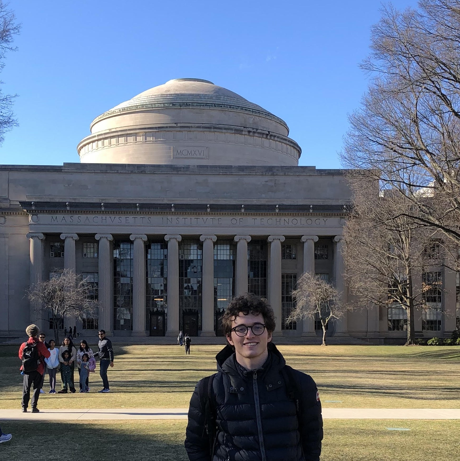
ANDREA BRIGLIADORI

Hello, my name is Andrea Brigliadori and I have recently submitted my thesis for the MSc in AI for Biomedicine and Healthcare at University College London. In this page, I present lines of research I would like to investigate, and some articles I have written.
Title
Date
This work focused on improving the ability of machine learning models to generalise across MRI scanner settings when estimating properties of brain microstructure. Microstructure refers to the tissue structure at the micrometre scale, and its properties are indicative of the health condition of the brain. The quantitative description of these properties can significantly accelerate the diagnosis of neurological disorders such as stroke and multiple sclerosis. Capturing information on brain microstructure non-invasively from living human beings has been made possible by an advanced magnetic resonance imaging technique known as diffusion-weighted MRI. Using two main scanner settings, known as b-vectors and b-values, this technique provides images that can be leveraged by computational models to estimate microstructural properties. There is growing interest in obtaining these estimates using machine learning models, since they offer great efficiency advantages compared to traditional methods. However, these models typically do not generalise well to changes of the scanner settings, and this limitation hampers their clinical utility. Recently, a geometric deep learning model known as spherical convolutional neural network (SCNN) has been shown to maintain its performance despite variations in the b-vectors. Nonetheless, the model provides no performance guarantees for b-values unseen during training. The aim of this project is to enhance the generalisability of SCNNs to new b-values. A novel SCNN architecture that accepts b-values as input is proposed and shown to improve performance on both seen and unseen b-values, although further hyperparameter tuning may be needed to obtain stronger evidence.
This work focuses on quantifying iron overload in the liver from magnetic resonance images. Measuring the amount of iron in the body is essential for the treatment of patients suffering from thalassemias, a class of widespread genetic diseases compromising the production of red blood cells. Without frequent blood transfusions, many of these patients do not reach the age of five. In turn, transfusions may lead to toxic accumulations of iron, potentially inducing severe health complications such as cardiac failure. In order to prevent this, it is critical to remove the excess iron in the body. At present, the only effective method is to use medications able to bind iron and assist in its excretion. However, it is necessary to tailor the length and intensity of this therapy to the specific patient, hence clinicians need to know the amount of surplus iron to remove. To approach this problem, computational models have been developed and applied on magnetic resonance images to estimate liver iron overload, which has been shown to closely relate to the total body iron content. In this study, we contribute to the field with an in-depth analysis of one of these methods, obtaining promising estimation results.
Full-text PDF